

Understanding how plasmalogens influence synaptic transmission is critical for advancing longevity science. Recent research indicates that plasmalogen deficiency can significantly alter membrane fluidity, which directly impacts the speed of synaptic vesicle fusion. According to studies published in the Journal of Lipid Research, membrane lipid composition is a primary determinant of vesicle dynamics. This guide outlines the scientific protocol for measuring these effects, bridging the gap between cellular biology and clinical longevity.
What Are Plasmalogens?
Plasmalogens are a unique class of ether phospholipids found in high concentrations in the brain, heart, and skeletal muscle. Unlike standard phospholipids, plasmalogens contain a vinyl ether bond at the sn-1 position of the glycerol backbone. This structural feature is not merely cosmetic; it provides critical protection against oxidative stress and regulates membrane fluidity.
Plasmalogens are specialized ether phospholipids essential for maintaining the structural integrity and functional efficiency of cell membranes. Their presence in synaptic vesicles is particularly important because the fusion process requires rapid changes in membrane curvature and lipid packing density. When plasmalogen levels decline, the membrane becomes less fluid and more rigid, potentially slowing down the release of neurotransmitters.
For a deeper understanding of their biological role, explore our comprehensive overview on What Are Plasmalogens. Additionally, the Critical Nutrients and Standards for Optimal Plasmalogen Synthesis article details the cofactors required for their biosynthesis.
The Synaptic Fusion Mechanism
To measure the effect of plasmalogens on fusion speed, one must first understand the mechanism of synaptic vesicle fusion. Synaptic vesicles are small, membrane-bound sacs that store neurotransmitters. When an action potential reaches the presynaptic terminal, these vesicles must fuse with the presynaptic membrane to release their contents into the synaptic cleft.
A synapse is the communication site where one nerve cell passes information to another cell via neurotransmitter release. This process is mediated by a protein complex known as the SNARE complex (Soluble N-ethylmaleimide-sensitive factor Attachment protein REceptor). The SNARE proteins pull the vesicle and plasma membranes together, overcoming the energy barrier required for fusion.
Plasmalogens influence this process in two key ways:
- Membrane Fluidity: Plasmalogens maintain optimal lipid order. If the membrane is too rigid, the SNARE complex cannot efficiently drive the curvature changes needed for fusion.
- Oxidative Protection: The vinyl ether bond acts as a sacrificial antioxidant, protecting the polyunsaturated fatty acids in the membrane from peroxidation. Oxidized lipids increase membrane stiffness, directly inhibiting fusion speed.
For more details on how these cellular structures function, read How The Synapse Works in the Brain & Body and Cell Membranes Explained.
Measurement Methodology
Measuring the direct effect of plasmalogens on synaptic vesicle fusion speed requires advanced electrophysiological and imaging techniques. This protocol is typically conducted in vitro using hippocampal or cortical neuron cultures, or in vivo using acute brain slices.
Step 1: Preparation of Neuronal Cultures
Begin by isolating primary neurons from rodent models. Divide the cultures into two groups: a control group with normal plasmalogen levels and an experimental group with induced plasmalogen deficiency. Deficiency can be modeled using genetic knockouts of the PEX (peroxisomal biogenesis) genes or by treating cells with specific inhibitors of plasmalogen synthesis.
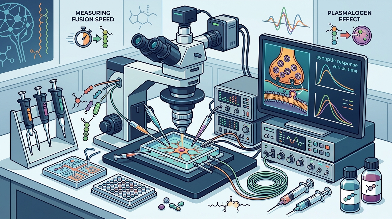
Step 2: Electrophysiological Recording
Use whole-cell patch-clamp electrophysiology to measure the kinetics of neurotransmitter release. Specifically, monitor the excitatory postsynaptic current (EPSC) amplitude and rise time. A slower rise time indicates delayed vesicle fusion. According to data from the National Center for Biotechnology Information, alterations in EPSC kinetics are a reliable proxy for changes in fusion pore dynamics.
Step 3: Fluorescent Imaging of Vesicle Dynamics
Employ pH-sensitive fluorescent dyes such as pHluorin-tagged synaptophysin. This allows for real-time visualization of vesicle fusion events. When a vesicle fuses, the acidic interior is exposed to the neutral extracellular pH, causing a rapid increase in fluorescence. By analyzing the frequency and amplitude of these fluorescence transients, researchers can quantify the speed and efficiency of fusion.
Step 4: Lipidomic Analysis
Concurrently, perform lipidomic profiling on the same neuronal samples. Use mass spectrometry to quantify the levels of specific plasmalogen species, such as PlasEtnPCho and PlasEtnPEtn. Correlate these lipid levels with the electrophysiological data. Studies suggest that a significant correlation exists between plasmalogen density and synaptic efficacy.
Advanced Testing & Biomarkers
While in vitro models provide mechanistic insights, advanced health measurement is changing how we understand these processes in living systems. For clinical applications, direct measurement of synaptic fusion speed is not feasible. Instead, we rely on biomarkers that reflect membrane integrity and synaptic health.
Advanced health measurement looks deeper than standard blood panels. It examines patterns in lipid biology, oxidative stress, and cellular energy. The goal is to understand how the body is functioning across systems, where early biological stress may be developing, and how membrane health impacts cognitive resilience.
Key biomarkers include:
- Plasmalogen Ratio: The ratio of plasmalogens to other phospholipids in red blood cells or plasma.
- Oxidative Stress Markers: Levels of F2-isoprostanes, which indicate lipid peroxidation.
- Inflammatory Markers: C-reactive protein (CRP) and interleukin-6 (IL-6), which can degrade membrane integrity.
For a comprehensive guide on these metrics, visit The Importance of Advanced Health Measurements in Health and Longevity. Additionally, Understanding Plasmalogen Levels provides insight into how to interpret these biomarkers in a clinical context.
Key Takeaways
- Plasmalogens are ether phospholipids that maintain membrane fluidity and protect against oxidative stress.
- Synaptic vesicle fusion speed is directly influenced by the lipid composition of the presynaptic membrane.
- Plasmalogen deficiency leads to increased membrane rigidity, slowing down neurotransmitter release.
- Electrophysiology and fluorescent imaging are the gold standards for measuring fusion kinetics in research.
- Advanced lipidomic testing can serve as a proxy for synaptic health in clinical settings.
- Oxidative stress is a primary driver of plasmalogen depletion and subsequent synaptic dysfunction.
- Longitudinal tracking of plasmalogen levels is more predictive of cognitive health than single-time testing.
Frequently Asked Questions
How do plasmalogens affect synaptic vesicle fusion?
Plasmalogens maintain the optimal fluidity of the synaptic membrane. By preventing excessive rigidity, they allow the SNARE protein complex to efficiently drive the membrane curvature required for vesicle fusion and neurotransmitter release.
What is the best way to measure plasmalogen levels?
Advanced lipidomic profiling using mass spectrometry is the most accurate method. This can be performed on plasma, red blood cells, or brain tissue samples to quantify specific plasmalogen species.
Can plasmalogen deficiency cause cognitive decline?
Yes. Research indicates that low plasmalogen levels are associated with impaired synaptic plasticity, reduced memory formation, and accelerated neurodegeneration. This is due to the increased oxidative damage and reduced membrane efficiency in the brain.
What are the signs of low plasmalogen levels?
Signs may include cognitive fog, reduced memory retention, increased oxidative stress markers, and decreased membrane integrity in blood tests. In severe cases, it can lead to neurological disorders.
How does oxidative stress impact plasmalogens?
Oxidative stress depletes plasmalogens because their vinyl ether bond acts as a sacrificial antioxidant. When plasmalogens neutralize free radicals, they are consumed, leading to a net decrease in their concentration within cell membranes.
Are there dietary sources of plasmalogens?
Plasmalogens are synthesized endogenously in the body. However, their production requires specific nutrients such as choline, methionine, and vitamins B12 and B6. Dietary support for these cofactors is essential for maintaining plasmalogen levels.
What is the difference between plasmalogens and other phospholipids?
Plasmalogens contain a vinyl ether bond at the sn-1 position, whereas other phospholipids contain an ester bond. This structural difference makes plasmalogens more resistant to oxidative damage and more effective at regulating membrane fluidity.
How can I support plasmalogen synthesis?
Supporting plasmalogen synthesis involves ensuring adequate intake of essential cofactors, reducing oxidative stress through lifestyle changes, and potentially using targeted supplements under medical guidance. Regular advanced health measurements can help monitor progress.
Optimize Your Synaptic Health Today
Understanding the link between plasmalogens and synaptic function is the first step toward preserving cognitive longevity. By monitoring your lipid profiles and supporting your body's natural biosynthesis pathways, you can protect your brain's most critical communication networks.
Ready to take control of your healthspan? Contact Plasmalogen Science today to schedule a consultation with our experts and learn how advanced testing can reveal your unique plasmalogen status.